
Ovary Diagrams to Print 101 Diagrams
T.S. of Mammalian Ovary. The mammalian ovaries are the reproductive organ in females inside which sex cells like eggs or ova are produced. In addition to the sex cells, the ovaries come in pairs and produce Oestrogen and Progesterone hormones that trigger reproduction in females. While ovaries are the primary reproductive organs in females, males have testis as their primary reproductive organ.

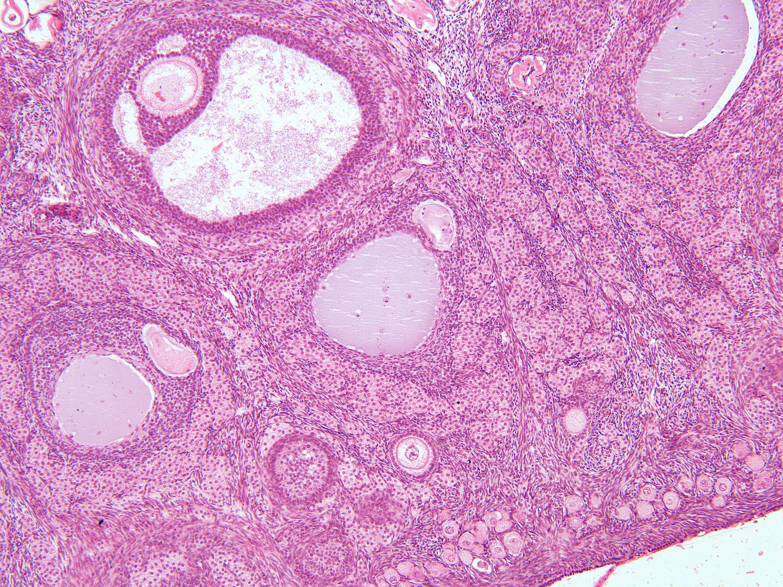
OvaryFollicles200x Ovaries, Reproductive system, Human development
Oogenesis in non-human mammals Diagram showing the reduction in number of the chromosomes in the process of maturation of the ovum. (In mammals, the first polar body normally disintegrates before dividing, so only two polar bodies are produced. [citation needed])In mammals, the first part of oogenesis starts in the germinal epithelium, which gives rise to the development of ovarian follicles.

Pin by Ali Mortza on histology Histology slides, Medical studies, Science equipment
Some species reproduce only once during their life span, whereas others, such as mammals, have reproductive cycles that are hormonally regulated; the female ovary is central to the process of reproduction and produces germ cells, as well as gonadal hormones. Keywords Corpus Luteum Follicular Fluid Zona Pellucida Meiotic Division Primordial Follicle

Ts of ovary Biology 12468417
Solution 1 Externally, each testis is covered by three layers. These are: a. Tunica vaginalis: It is the outermost incomplete peritoneal covering made up of connective tissue and epithelium. b. Tunica albuginea: It is the middle layer formed by fibrous connective tissue. c. Tunica vasculosa:
Ovaries Function, Location, Hormones Produced. What control it?
Abstract. The prevailing concept of ovarian morphology, at the turn of the century, was that the important functional components were the follicles, especially the oocyte, and the corpora lutea. If one scans the voluminous literature that has accumulated during the past 45 years, one will find that the follicles and the corpora lutea still.

Mammalian ovary and graafian follicles, light micrograph Stock Image C047/6173 Science
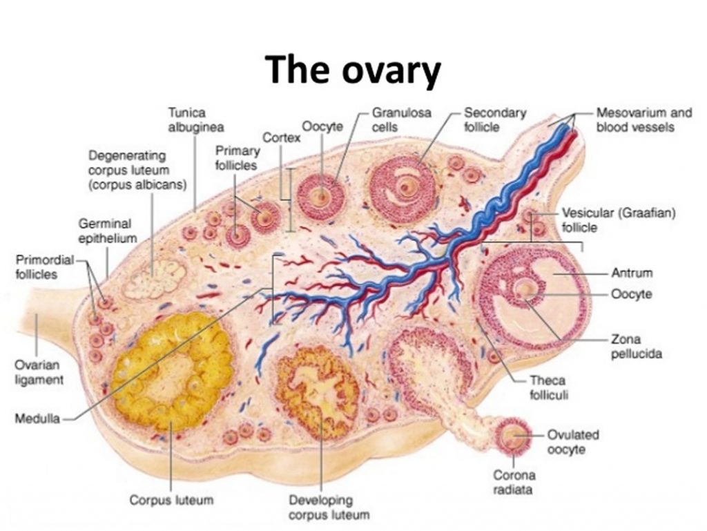
T.S. of Mammalian Ovary A mammalian ovary is a solid structure bounded germinal epithelium followed by a thick layer of fibrous tissue called tunica albuginea. The ovary consists of the outer cortex and inner medulla. The medulla consists many rounded or oval bodies called ovarian follicles at various stages of development.

T.S. of ovary Ovaries, Happy independence, Save
Both functions of the mammalian ovary, the endocrine and (synthesis and secretion of steroid hormones) and exocrine (production of ova), depend upon the presence and cyclic growth of follicles, as the depletion of primordial follicles from the ovary leads to cessation of these f-unctions or female reproduction in mammals, or to postmenopausal period in humans.

How to draw T.S. of ovary । structure of mammalian ovary । Biology class 12 YouTube
Although the testis and ovary are functionally analogous and arise from a common primordial structure, the genital ridge, they are remarkably different organs, and their development is driven by distinct programs of gene regulation and cellular organization.
Ovarian Follicles Slide
Ovary Structure and Function. The ovary is a female reproductive organ that contains an egg called an ovum. This travels down the fallopian tube further into the after being released, where it could be fertilized by sperm. On either side of the body, there is an ovary (from Latin ovarium 'egg, nut'). Hormones that play a part in the menstrual.
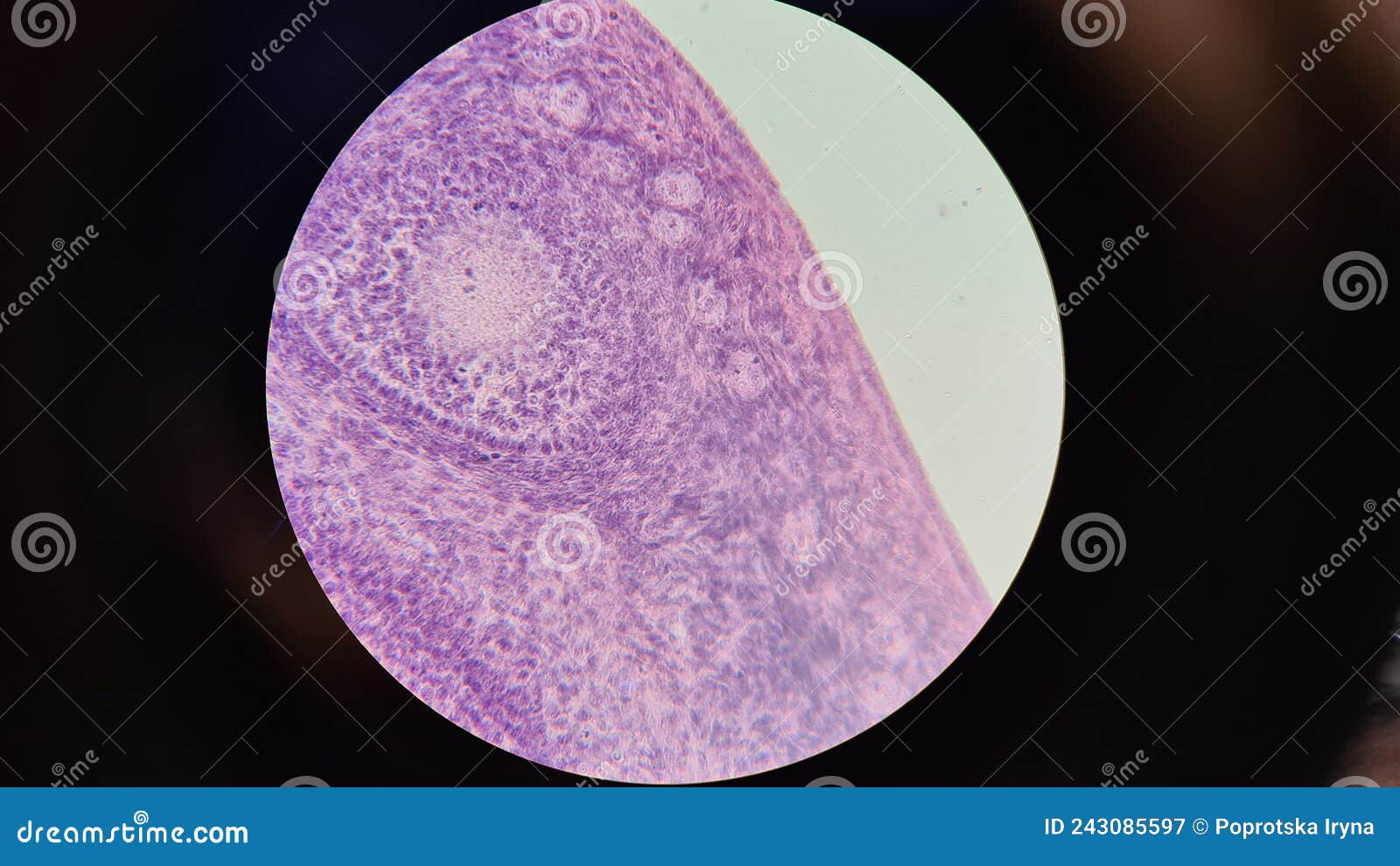
Mammalian Ovary Under A Microscope Stock Image 243085597
In mammals, the blastula forms the blastocyst in the next stage of development. Here the cells in the blastula arrange themselves in two layers: the inner cell mass and an outer layer called the trophoblast. The inner cell mass is also known as the embryoblast; this mass of cells will go on to form the embryo..
[Solved] Mammal Ovary Follicles Need Labeling Primary follicle Secondary... Course Hero
Most mammals, including female humans, possess two ovaries, right and left. The development of gametes inside ovaries is called oogenesis. TS of mammalian ovaries will help you to study the several components of ovaries. Observation. There is a mass of tissue lined with germinal epithelium in the ovarian region.
ANIMAL ANATOMY BIOLOGY4ISC
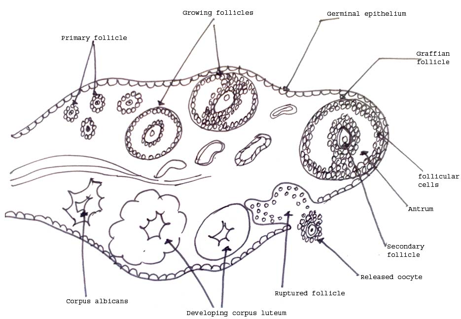
1. An ovary is a germinal epithelium bounded by a solid structure covered by a thick layer of. fibrous tissue known as tunica albuginea. 2. It consists of an inner medulla and an outer cortex. 3. The medulla comprises several round or oval bodies known as ovarian follicles. 4. Follicle development takes place in the following stages:

TS of ovary, demonstrating stage II of maturity. Showing type three of... Download Scientific
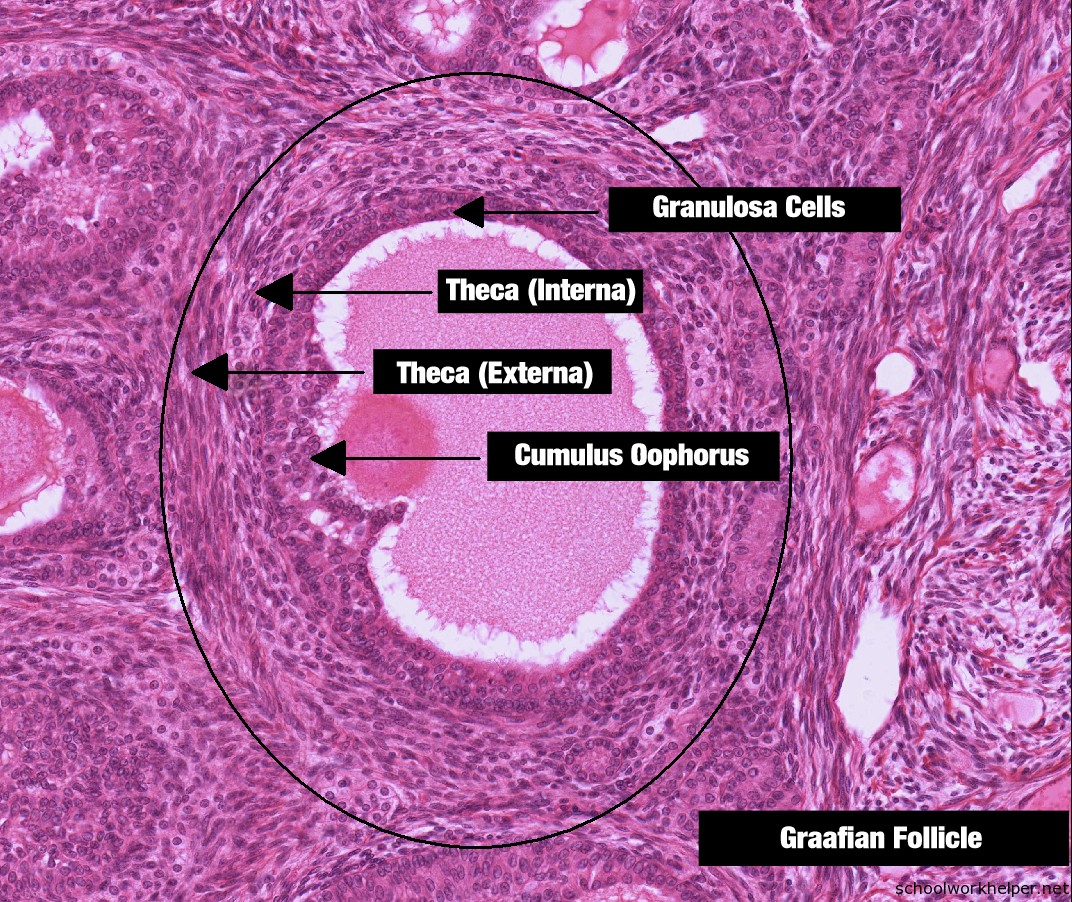
T.S. of Ovary (i) In the section of ovary, there is a mass of tissue lined with germinal epithelium. Inside that you will Graafian Follicle observe an ovum, which is a cell surrounded by one to several layers of follicular cells. As the ovum matures, the number of surrounding follicular cell layer increases (Fig. 4.2).

Ovario Normal, Desarrollo Folicular Y Ovulación Stock de ilustración Ilustración de normal
1. The medulla 2. The cortex Medulla The medulla, also known as the zona vasculosa, is the deeper core region of the ovary. It has a stroma made up of loose connective tissues. Near the hilum, it comprises blood arteries, lymphatics, nerve fibres, and bundles of smooth muscle fibres. Cortex
Ovary from cat (100 X)
The ovary has an outer cortex and an inner medulla. The T.S.of the mammalian ovary reveals the rounded oval bodies, also known as ovarian follicles. The follicles develop from stage 1 to Graffian follicles to corpus luteum. The development of the follicles is the necessary study of the transverse section of an ovary. Study of T.S. of Mammalian.

Professor Olexik Ovaries, Human anatomy and physiology, Reproductive system
5 - The ovary. Published online by Cambridge University Press: 05 June 2012. In the mature animal the structure and function of the ovary is continually changing. Gonadotrophins secreted by the anterior pituitary gland stimulate the growth of Graafian follicles (folliculogenesis), ovulation, and the formation of corpora lutea.
